PE anti |
您所在的位置:网站首页 › egfr1 › PE anti |
PE anti
Pricing & Availability
Product Details
Antigen Details
Documentation
Reviews
Related Protocols
Related Products
Related Pages & Pathways
Related FAQs
Other Formats
Concentration & Expiration Lookup
Certificate of Analysis
Pricing & Availability
Clone
AY13 (See other available formats)
Regulatory Status
RUO
Other Names
Proto-oncogene c-ErbB-1, Receptor tyrosine-protein kinase erbB-1, HER1
Isotype
Mouse IgG1, κ
Ave. Rating
Submit a Review
Product Citations
publications
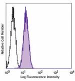
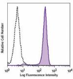
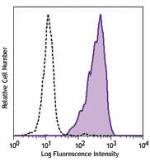
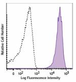
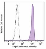
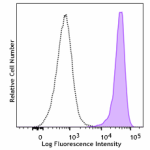
 Human cervical cancer cell line HELA was stained with EGFR (clone AY13) PE (filled histogram) or mouse IgG1, κ PE isotype control (open histogram).
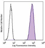
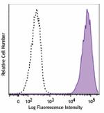
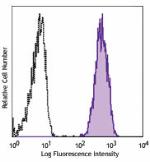
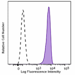
 Human cervical cancer cell line HELA was stained with EGFR (clone AY13) PE (filled histogram) or mouse IgG1, κ PE isotype control (open histogram).
Compare all formats
See PE spectral data
Cat #
Size
Price
Quantity
Check Availability
Save
Input string was not in a correct format.
352903
25 tests
£81
Input string was not in a correct format.
352904
100 tests
£221
Description
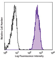
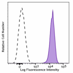
Human cervical cancer cell line HELA was stained with EGFR (clone AY13) PE (filled histogram) or mouse IgG1, κ PE isotype control (open histogram).
Compare all formats
See PE spectral data
Cat #
Size
Price
Quantity
Check Availability
Save
Input string was not in a correct format.
352903
25 tests
£81
Input string was not in a correct format.
352904
100 tests
£221
Description
Epidermal growth factor receptor (EGFR) is a transmembrane glycoprotein and member of the protein kinase superfamily that regulates cell growth and differentiation. EGFR binds EGF, TGF-α, amphiregulin, betacellulin, heparin-binding EGF-like growth factor, GP30, and vaccinia virus growth factor - all members of the EGF family. Ligand binding induces EGFR dimerization and autophosphorylation, initiating the MAPK, Akt, and JNK signaling pathways. EGFR is expressed by epithelial and endothelial cells and is frequently expressed by epithelial carcinomas. Product Details Technical Data Sheet (pdf) Product Details Verified Reactivity Human Antibody Type Monoclonal Host Species Mouse Immunogen Non-small cell lung cancer (NSCLC) cell line NCI-H322 Formulation Phosphate-buffered solution, pH 7.2, containing 0.09% sodium azide and BSA (origin USA) Preparation The antibody was purified by affinity chromatography and conjugated with PE under optimal conditions. Concentration Lot-specific (to obtain lot-specific concentration, please enter the lot number in our Concentration and Expiration Lookup or Certificate of Analysis online tools.) Storage & Handling The antibody solution should be stored undiluted between 2°C and 8°C, and protected from prolonged exposure to light. Do not freeze. ApplicationFC - Quality tested Recommended UsageEach lot of this antibody is quality control tested by immunofluorescent staining with flow cytometric analysis. For flow cytometric staining, the suggested use of this reagent is 5 µl per million cells in 100 µl staining volume or 5 µl per 100 µl of whole blood. Excitation Laser Blue Laser (488 nm)Green Laser (532 nm)/Yellow-Green Laser (561 nm) Application References(PubMed link indicates BioLegend citation) Yamaguchi M, et al. 2009. The 15th Annual Meeting Japan Society of Gene Therapy. p1056. Abstract 92. Product Citations Wang Y, et al. 2014. Biomaterials. 35:4297. PubMed Sreevalsan S, et al. 2020. EMBO Rep. 21:e50155. PubMed Kitamura Y, et al. 2021. Sci Adv. 7: . PubMed Halim L, et al. 2017. Cell Rep. 10.1016/j.celrep.2017.06.079. PubMed Bak R and Proteus M. 2017. Cell Rep. 10.1016/j.celrep.2017.06.064. PubMed Wang JL, et al. 2022. Cancers (Basel). 14:. PubMed Lu T, et al. 2022. Nat Commun. 13:2576. PubMed Jayasinghe MK, et al. 2022. Theranostics. 12:3288. PubMed Aldeghaither DS, et al. 2019. Cancer Immunol Res. 7:230. PubMed Carnevale J, et al. 2022. Nature. 609:174. PubMed Grass G, et al. 2013. J Biol Chem. 288:26089. PubMed Ahn S, et al. 2019. Cancer Immunol Res. 0.828472222. PubMed Edinger N, et al. 2016. PLoS One. 11: 0162321. PubMed Wing A, et al. 2018. Cancer Immunol Res. 6:605. PubMed Okada R, et al. 2021. EBioMedicine. 67:103345. PubMed Han X, et al. 2017. Mol Ther. 10.1016/j.ymthe.2017.07.009. PubMed Brohl AS, et al. 2021. Cell Rep. 37:110047. PubMed Jarantow S, et al. 2015. J Biol Chem. 290: 24689 - 24704. PubMed Li G, et al. 2021. Mol Ther Oncolytics. 22:507. PubMed Fischer A, et al. 2020. Toxins (Basel). 12:00. PubMed Quijano-Rubio A, et al. 2022. Nat Biotechnol. Online ahead of print. PubMed RRID AB_10898161 (BioLegend Cat. No. 352903) AB_10896794 (BioLegend Cat. No. 352904) Antigen Details Structure Member of the protein kinase superfamily; transmembrane glycoprotein; ligand binding induces dimerization and autophosphorylation DistributionEpithelial cells and endothelial cells; frequently found in epithelial carcinomas Function Controls cell growth and differentiation Interaction MAPK, Akt, JNK Ligand/Receptor Members of the epidermal growth factor (EGF) family such as EGF, TGF-α, amphiregulin, betacellulin, heparin-binding EGF-like growth factor, GP30 and vaccinia virus growth factor Cell Type Endothelial cells, Epithelial cells Biology Area Cell Biology, Cell Cycle/DNA Replication, Immunology, Innate Immunity, Neuroscience, Synaptic Biology Molecular Family CD Molecules, Growth Factors Antigen References1. da Cunha Santos G, et al. 2011. Annu. Rev. Pathol. 6:49. 2. Gusterson BA and Hunter KD. 2009. Lancet Oncol. 10:522. 3. Mano M and Humblet Y. 2008. Nat. Clin. Pract. Oncol. 5:415. 4. Pao W and Chmielecki J. 2010. Nat. Rev. Cancer 10:760. Gene ID 1956 View all products for this Gene ID Specificity (DOES NOT SHOW ON TDS): EGFR Specificity Alt (DOES NOT SHOW ON TDS): EGFR UniProt View information about EGFR on UniProt.org Documentation Technical Data Sheet (pdf) Certificate of Analysis Safety Data Sheet Reviews Review This Product Related Protocols Cell Surface Flow Cytometry Staining Protocol Related Products Description Clone Applications Related Pages & Pathways Pathways EGF Pathway ErbB Family Pathway ERK Signaling FAK1 Pathway p38 Signaling Pages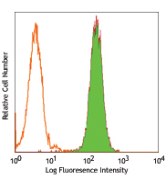 APC anti-human EGFR
Clone:
AY13
APC anti-human EGFR
Clone:
AY13
 Brilliant Violet 711™ anti-human CD25
Clone:
M-A251
Brilliant Violet 711™ anti-human CD25
Clone:
M-A251
 Brilliant Violet 605™ anti-human CD127 (IL-7Rα)
Clone:
A019D5
Brilliant Violet 605™ anti-human CD127 (IL-7Rα)
Clone:
A019D5
 Go To Top
Version: 1 Revision Date: 11/30/2012
Go To Top
Version: 1 Revision Date: 11/30/2012
For research use only. Not for diagnostic use. Not for resale. BioLegend will not be held responsible for patent infringement or other violations that may occur with the use of our products.
*These products may be covered by one or more Limited Use Label Licenses (see the BioLegend Catalog or our website, www.biolegend.com/ordering#license). BioLegend products may not be transferred to third parties, resold, modified for resale, or used to manufacture commercial products, reverse engineer functionally similar materials, or to provide a service to third parties without written approval of BioLegend. By use of these products you accept the terms and conditions of all applicable Limited Use Label Licenses. Unless otherwise indicated, these products are for research use only and are not intended for human or animal diagnostic, therapeutic or commercial use.
8999 BioLegend Way, San Diego, CA 92121 www.biolegend.com Toll-Free Phone: 1-877-Bio-Legend (246-5343) Phone: (858) 768-5800 Fax: (877) 455-9587 Compare Data Across All FormatsThis data display is provided for general comparisons between formats. Your actual data may vary due to variations in samples, target cells, instruments and their settings, staining conditions, and other factors. If you need assistance with selecting the best format contact our expert technical support team. Purified anti-human EGFR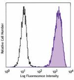
 PE anti-human EGFR
 APC anti-human EGFR
 Alexa Fluor® 488 anti-human EGFR
 Brilliant Violet 421™ anti-human EGFR
 PE/Cyanine7 anti-human EGFR
 PerCP/Cyanine5.5 anti-human EGFR
 Alexa Fluor® 594 anti-human EGFR
 Alexa Fluor® 647 anti-human EGFR
 Brilliant Violet 711™ anti-human EGFR
 PE/Dazzle™ 594 anti-human EGFR
 TotalSeq™-A0132 anti-human EGFR
Brilliant Violet 605™ anti-human EGFR
TotalSeq™-A0132 anti-human EGFR
Brilliant Violet 605™ anti-human EGFR
 APC/Fire™ 750 anti-human EGFR
 TotalSeq™-B0132 anti-human EGFR
TotalSeq™-C0132 anti-human EGFR
Biotin anti-human EGFR
TotalSeq™-B0132 anti-human EGFR
TotalSeq™-C0132 anti-human EGFR
Biotin anti-human EGFR
|
【本文地址】
今日新闻 |
推荐新闻 |